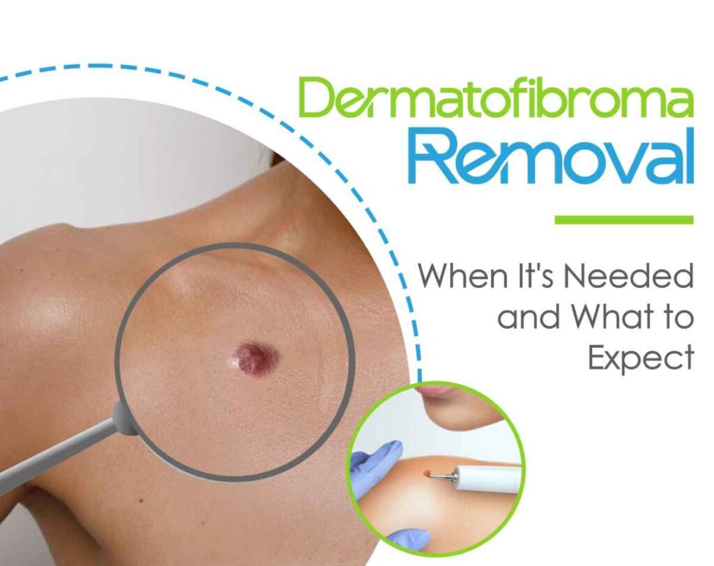
dermatofibroma
Discovering a new bump on your skin can naturally cause concern. If you have noticed a small, firm, brownish nodule often on your lower legs or arms that feels like a tiny pebble beneath the surface, you may be dealing with a dermatofibroma.
While these common skin growths are completely benign (non-cancerous), their appearance can sometimes mimic more serious skin conditions like melanoma. Understanding what a dermatofibroma is, why it forms, and when to consider professional treatment is essential for maintaining your skin health and peace of mind.
In this comprehensive guide, we will explore the characteristics of dermatofibromas, how dermatologists diagnose them, the primary methods for dermatofibroma removal, and what to expect during recovery.
What is a Dermatofibroma?
A dermatofibroma (also known as a benign fibrous histiocytoma) is a common, non-cancerous skin growth made up of an overgrowth of fibrous tissue, blood vessels, and immune cells within the dermal layer of the skin.
These benign nodules are typically harmless and do not transform into skin cancer. However, because they sit deep within the dermis, they rarely disappear on their own and can remain permanently unless surgically treated.
Common Physical Characteristics
- Appearance: Small, firm bumps ranging from pink or reddish-brown to dark brown or purple.
- Size: Usually small, typically measuring between 3 to 10 millimeters in diameter.
- Location: Most frequently found on the lower legs, arms, and upper back, though they can develop anywhere on the body.
- The “Dimple Sign”: A classic physical feature of a dermatofibroma is that when you pinch the skin around the bump, it dimples inward rather than pushing outward.
- Sensory Symptoms: While usually asymptomatic, some dermatofibromas may occasionally itch, feel tender, or cause discomfort if rubbed by clothing or shaved over.
What Causes a Dermatofibroma to Develop?
The exact cause of dermatofibromas is not entirely clear, but dermatologists generally consider them a reactive tissue response rather than a true neoplastic tumor.
Common triggering factors include:
- Minor Skin Trauma: Many dermatofibromas appear following a minor injury to the skin, such as a bug bite, a splinter, or a nick from a razor blade.
- Insect Bites: Reactions to mosquito or spider bites frequently trigger localized inflammation that can develop into fibrous tissue.
- Gender Predisposition: Dermatofibromas occur significantly more often in women than in men, particularly during young adulthood and middle age.
- Immune Responses: In rare instances, individuals with compromised immune systems may develop multiple dermatofibromas across different areas of the body.
Diagnosing a Dermatofibroma: Why Professional Evaluation Matters
Before considering any dermatofibroma removal treatment, obtaining an accurate medical diagnosis from a qualified dermatologist or skin cancer specialist is essential.
Because dermatofibromas contain dark pigment, they can occasionally resemble dermatological conditions such as dermatofibrosarcoma protuberans (DFSP), basal cell carcinoma, or cutaneous melanoma.
How Doctors Confirm a Dermatofibroma
- Dermoscopy: A clinician uses a specialized magnifying light called a dermatoscope to evaluate the pigment pattern. A classic dermatofibroma typically displays a central white patch surrounded by a fine pigment network.
- Physical Examination: Performing the “pinch test” to check for the characteristic inward dimple.
- Skin Biopsy: If the lesion displays atypical features, irregular borders, or rapid growth, your doctor will perform a punch or shave biopsy to confirm its benign nature under a microscope.
The Dermatofibroma Removal Process: Medical and Surgical Options
Because dermatofibromas are harmless, removal is generally optional. Most patients seek dermatofibroma removal for cosmetic reasons, if the nodule is located in an inconvenient area subject to constant friction, or if it causes persistent itching or pain.
Because these growths extend deep into the dermal layer, complete removal requires specific clinical procedures.
1. Surgical Excision (The Gold Standard)
Full surgical excision is the most effective and definitive treatment for a dermatofibroma.
- How It Works: After numbing the area with local anesthesia, the surgeon uses a scalpel to remove the entire lesion, including its deep dermal roots, along with a tiny margin of surrounding skin. The resulting wound is closed with surgical sutures.
- Pros: Guarantees 100% complete removal of the nodule with virtually zero chance of recurrence.
- Cons: Because the incision cuts through the full thickness of the skin, excision will always leave a permanent, linear scar. In some cases, the resulting scar may be more noticeable than the original bump.
2. Cryotherapy (Liquid Nitrogen Freezing)
Cryotherapy is a minimally invasive alternative that uses extreme cold to target the surface tissue.
- How It Works: Liquid nitrogen is applied directly to the dermatofibroma to freeze and destroy the superficial cells. Over the following weeks, the treated tissue blisters and sloughs off.
- Pros: Quick, requires no scalpel or stitches, and carries minimal recovery downtime.
- Cons: Cryotherapy usually only flattens the top portion of the growth rather than removing the deep core. The bump may gradually regrow over time, and localized pigment loss (hypopigmentation) can occur.
3. Shave Excision / Laser Therapy
- Shave Excision: The doctor uses a surgical blade to slice off the top raised portion level with the surrounding skin. While it smooths the skin surface, the deeper root remains, making regrowth common.
- Laser Treatment: Certain vascular or pigment lasers can reduce redness or hyperpigmentation associated with the lesion, but they do not eliminate the underlying fibrous mass.
Post-Removal Recovery and Wound Care
Proper post-procedure care is crucial for minimizing scarring and preventing infection following a dermatofibroma removal procedure.
- Keep the Site Clean and Dry: Follow your doctor’s instructions regarding bandage changes, typically keeping the area clean and covered for the first 24 to 48 hours.
- Apply Ointment: Using a petroleum-based ointment can promote optimal moist wound healing and minimize heavy scabbing.
- Limit Tension: Avoid strenuous activities or stretching that could put tension on the stitches, especially if the removal was on a limb or joint.
- Sun Protection: Once healed, apply a high-SPF sunscreen over the new scar tissue to prevent hyperpigmentation.
Frequently Asked Questions
Can a dermatofibroma turn into skin cancer?
No. A dermatofibroma is a strictly benign skin growth and does not transform into melanoma or non-melanoma skin cancers. However, any new or changing skin spot should be evaluated by a healthcare professional to confirm an accurate diagnosis.
Can I remove a dermatofibroma at home?
Never attempt to cut, squeeze, or freeze a dermatofibroma at home. Because these nodules extend deep into the dermis, DIY removal attempts can lead to severe infections, excessive bleeding, permanent scarring, and incomplete removal.
Will dermatofibroma removal leave a scar?
Yes. Because a dermatofibroma resides deep in the skin layers, effective removal methods particularly surgical excision will leave a permanent scar. A skilled practitioner will align the incision with your skin’s natural tension lines to minimize scar visibility.
Do dermatofibromas grow back after removal?
If the nodule is fully excised surgically down through the dermal layer, recurrence is extremely rare. However, if partial removal methods like shave excision or cryotherapy are used, the remaining deep tissue may eventually cause the bump to regrow.
Protecting Your Skin Health
While a dermatofibroma is a harmless skin condition, experiencing changes in your skin’s texture, color, or shape always warrants a professional medical opinion. Choosing whether to undergo dermatofibroma removal comes down to balancing cosmetic preference against the likelihood of a small surgical scar.
Consulting an experienced clinician ensures an accurate diagnosis, peace of mind, and access to safe, precise treatment options tailored to your needs.